
Heart Disease Testing & Diagnosis
At UM Capital Region Heart and Vascular Institute, our cardiologists have access to the latest technology and equipment for diagnostics and cardiac imaging.
When treating heart disease, an accurate diagnosis is an important first step to successful treatment.
Depending on your symptoms, our team may suggest a number of different diagnostic options to allow us to examine your heart and find the problem.
Cardiac Catheterizations
An interventional cardiologist will access your heart through a tiny tube that is inserted either through your groin, neck or arm. This tube is directed to the heart, and the interventional cardiologist will feed a tiny camera through that tube to your heart, allowing us to see exactly what is happening.
Cardiac catheterizations can be used to diagnose a heart issue, to take a biopsy, or to perform procedures.
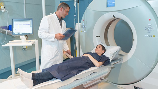
Chest CT
Computed tomography (CT) is a special X-ray that takes images from many different angles to build a complete visual of the heart without operating. With a chest CT, we focus the images on the area around the heart to allow us to see the full function of your heart.
Heart Scans
A heart scan, also know as a coronary calcium scan, is a special X-ray that helps detect plaque in the arteries. Plaque in the arteries can lead to a number of issues, including coronary artery disease and heart attacks.
Echocardiogram
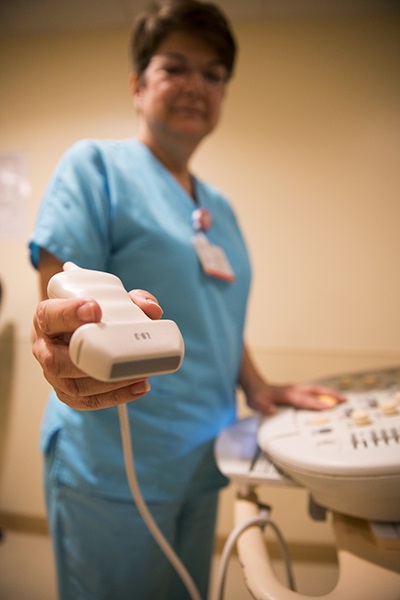
An echocardiogram, also called cardiac echo, is an ultrasound of the heart that allows our team to see your heart beating and pumping blood in real time.
This helps us see if there are problems with the valves or chambers or detect congenital heart defects.
Electrocardiogram (ECG or EKG)
An electrocardiogram allows our team to see the electrical signals in your heart. Many smart watches even have this as a function. We may ask you to wear a device, like a Holter monitor, for a day or several days to give us data on how your heart's electrical signals work over a period of time. This can help us to diagnose arrhythmias.
Exercise Stress Test
Exercise stress tests show us how your heart performs when it's working hard.
During your stress test, our team will monitor your heart's rate and electrical signals, along with your breathing and blood pressure, while you walk on a treadmill. We'll slowly increase the speed and incline, making you and your heart work harder.
We will review the results to help us make a diagnosis and recommendations for lifestyle changes.
Nuclear Stress Test
This test uses the same concepts as the exercise stress test with an added component: we inject you with a radioactive dye and use an imaging machine to create pictures showing your heart's function during the stress test.
We'll take images of your heart while at rest and while exercising, then compare the images. This method allows us to better tell if you're at risk for a heart attack.