
UM Multiple Sclerosis Neuroimaging Biomarker Research Group's Ongoing Research
7T MRI
The high-resolution images this technology captures provide further insights into MS pathology, including new biomarkers.
In collaboration with The F.M. Kirby Center for Functional Neuroimaging at the Kennedy Krieger Institute (KKI), we visualized cortical lesions (see Figure 1), lesions in the thalamus, lesions with quantitative susceptibility (QSM) rims suggestive of chronic inflammation and contrast enhancement suggestive of meningeal inflammation (see Figure 2).
As we continue this work, we hope to perfect these techniques and link our findings with clinical data, OCT data and blood/cerebrospinal fluid biomarkers.
UM’s Multiple Sclerosis Neuroimaging and Biomarker Research Group has multiple, ongoing projects related to the use of 7T MRI in MS, including two NIH/NINDS funded grants:
- 1R01NS122980-01: Pooled analysis of multiple sclerosis findings on multi-site 7 Tesla MRI
- 1R01NS104403-01: In vivo assessment of meningeal inflammation and its clinical impact in multiple sclerosis by 7 Tesla MRI
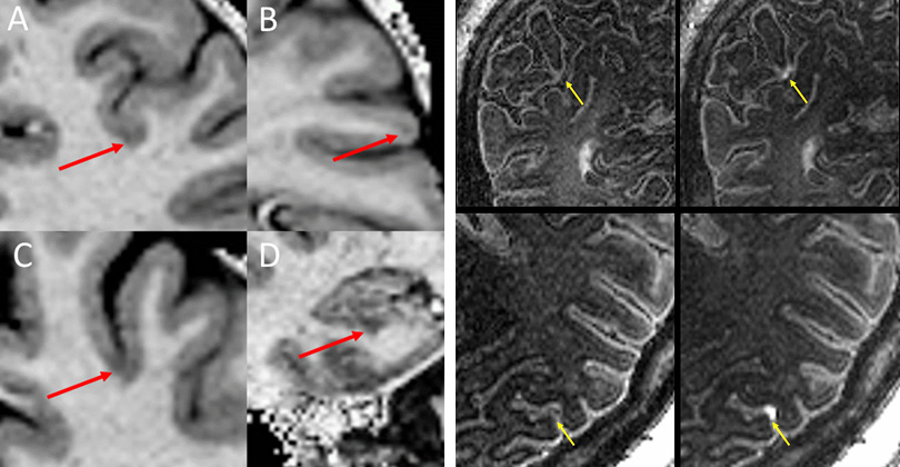
Figure 1 (left): Examples of cortical lesion subtypes. Cortical lesions were identified as hypointensities on T1w MP2RAGE images. Red arrows indicate lesion location. A = Leukocortical, B = Intracortical, C = Subpial, D = Hippocampal
Figure 2 (right): Meningeal contrast enhancement. Shown are pre-(left) and post-(right) contrast images from MPFLAIR at 7T. Contrast was seen in the meningeal space.
3T MRI
Together with colleagues in the Magnetic Resonance Research Center (MRRC) at the University of Maryland, we are working toward the development of new metabolic techniques and diffusion-based sequences using this technology.
Our research aims to find methods for quantifying astroglial changes related to MS and its neurodegenerative changes, in addition to measuring changes in metabolism in MS lesions.
Image Analysis Techniques
In research funded by the National MS Society, we are working with UM’s Department of Diagnostic Radiology and Nuclear Medicine to develop new image analysis techniques. This includes the use of machine-learning algorithms for the identification of lesions in the cortex of MS patients.
Through agreements with Biogen and the Cleveland Clinic Foundation, Dr. Harrison’s lab also has obtained access to MRI images from multiple clinical trials in MS. These are being used to train deep learning algorithms for predicting disability worsening and treatment response. This work is funded through a grant from the National MS Society:
- RG-2110-38460: Development of a Convolutional Neural Network for MRI Prediction of Progression and Treatment Response in Progressive Forms of Multiple Sclerosis
- Donate to Multiple Sclerosis Research
Adaptive Optics
In collaboration with scientists at the Food and Drug Administration, Dr. Harrison’s lab is also piloting the use of Adaptive Optics retinal imaging for multiple sclerosis. This technology is capable of imaging individual cells in the human retina, which may allow for more accurate monitoring of the effects of treatment on MS in the future. This research is funded by the Department of Defense:
- MS210103: Adaptive Optics Retinal Imaging in Multiple Sclerosis