
Xenogen Instrument Shared Service
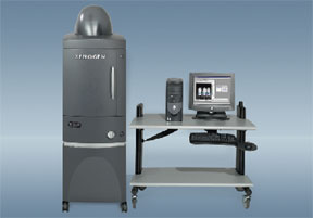 The Xenogen IVIS-200 System is capable of imaging bioluminescence and fluorescence
in living animals. A light-tight imaging chamber is coupled to a highly-sensitive
CCD camera system cooled to -95°C. This camera system is capable of quantitating
single-photon signals originating within the tissue of living mice. Up to five
mice can be imaged simultaneously and an integrated isoflurane gas manifold
allows rapid and temporary anesthesia of mice for imaging.
The Xenogen IVIS-200 System is capable of imaging bioluminescence and fluorescence
in living animals. A light-tight imaging chamber is coupled to a highly-sensitive
CCD camera system cooled to -95°C. This camera system is capable of quantitating
single-photon signals originating within the tissue of living mice. Up to five
mice can be imaged simultaneously and an integrated isoflurane gas manifold
allows rapid and temporary anesthesia of mice for imaging.
Bioluminescence from cell lines expressing firefly luciferase remains the most sensitive method for optical detection. The emission spectrum of luciferase at 37°C is largely above 600nm, and penetrates tissue very efficiently. Since excitation light is not required for luciferase bioluminescence, there is also minimal autofluorescence. For investigators interested in establishing luciferase-expressing cell lines for imaging, we recommend the pGL4 luciferase expression vector from Promega. The pGL4 plasmid is optimized for mammalian codon usage and routinely yields higher luciferase activities than other luciferase expression vectors.
 The IVIS-200 is also fully capable of imaging fluorescent signals, although
it is important to recognize the physical limitations of fluorescent imaging
in tissue. Both excitation and emission light below 600nm wavelength are efficiently
absorbed by tissue and generally do not penetrate more than a few microns. For
this reason, existing fluorescent proteins (GFP, YFP, DsRed etc.) are really
only useful for surface or subcutaneous imaging. Near-infrared fluorescent proteins
are currently a subject of intense study, so progress is expected soon. Near-infrared
fluorescent dyes are available (Cy5.5, ICG) and are well-established for in
vivo imaging applications.
The IVIS-200 is also fully capable of imaging fluorescent signals, although
it is important to recognize the physical limitations of fluorescent imaging
in tissue. Both excitation and emission light below 600nm wavelength are efficiently
absorbed by tissue and generally do not penetrate more than a few microns. For
this reason, existing fluorescent proteins (GFP, YFP, DsRed etc.) are really
only useful for surface or subcutaneous imaging. Near-infrared fluorescent proteins
are currently a subject of intense study, so progress is expected soon. Near-infrared
fluorescent dyes are available (Cy5.5, ICG) and are well-established for in
vivo imaging applications.
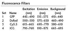
For those interested in developing fluorescent applications, the fluorescence filter sets available on the IVIS-200 are as follows:
For further information on developing in vivo imaging techniques for the IVIS-200, contact:
Stuart S. Martin, Ph.D.
Assistant Professor of Physiology
University of Maryland Greenebaum Comprehensive Cancer Center
