
X-ray
An X-ray is a painless medical test that makes images of bone and tissue, including solid organs like the lungs.
X-rays were discovered in 1895. X-ray imaging is the oldest, most common form of medical imaging.
A type of electromagnetic radiation, X-rays are similar to visible light.
However, X-rays have more energy and pass through many kinds of objects.
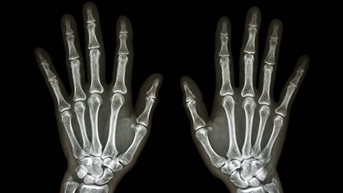
X-rays pass through your body to a medium, such as film or digital X-ray sensor, and make an image called a radiograph, which is much like a photograph. X-rays allow doctors to see and diagnose medical conditions and disorders, such as bone fractures and tumors.
Dense tissue, like bones and organs, allow less radiation to pass through and appear white or almost white while showing damage like fractures and tumors in great detail. X-rays pass right through soft tissue, like muscle, and make hardly any image. or just a vague outline or shadow.
You can go to any of our imaging locations for an X-ray.
Why You Need an X-ray
The most familiar use of X-ray is checking for broken bones. X-ray images can also show tumors and other abnormal masses, diseases like pneumonia and tuberculosis, as well as foreign objects and dental conditions.
What to Expect During an X-ray
You may lie on a table or stand against a wall, depending on your condition. The X-ray technologist will put the part of your body being X-rayed between the film or sensor and the X-ray machine. The technologist will ask you to hold still for the few seconds the exposure takes.
You may wear a lead apron during your X-ray to protect certain parts of your body, although the amount of radiation you get from an X-ray is small. It’s about the same dose you get naturally from the environment in about ten days.
Depending on why you have an X-ray, we may inject a contrast material or dye into a joint. Called an arthrogram, this procedure helps outline soft tissue structures in the joint. It may also confirm needle placement in the joint when fluid is removed or medication is injected into the joint.
Other Types of X-ray Imaging
At UMMC, we use other X-ray imaging technologies that include:
- CT (computed tomography), a painless, noninvasive, computerized diagnostic X-ray imaging test. CT scans create highly detailed, cross-sectional images, or slices, of internal organs, bones, soft tissue and blood vessels in color and 3D.
- Fluoroscopy is a noninvasive test that makes real-time, moving X-ray images using a continuous beam of radiation used for diagnosing internal body functions.
- Mammography is a specialized form of X-ray for breast imaging. Like X-ray, mammography now uses digital technology instead of film. Digital mammography provides higher quality imaging and shorter processing times, and can detect tumors too small to feel by hand.
- Intravenous pyelogram (IVP) is an X-ray examination of the kidneys, ureters and urinary bladder that uses intravenous contrast material.
Schedule an Appointment
To make an X-ray appointment, call 410-328-3225.